X-rays are basically the same thing as visible rays of light, it’s just that you just can’t see them. And just like visible light rays, X-rays do one of two things: either they pass through stuff, or stuff gets in the way of them. So, when you shoot X-rays at a horse some of the X-rays pass right through the horse, whereas some of the X-rays get stopped by tissues in the horse, especially by dense structures like bone. Historically these X-rays were captured on film, which then had to be brought back to the clinic, and developed in a dark room with special chemicals. Recently, digital devices were developed that capture the X-rays with digital X-ray sensors, instead of film. In order to obtain the image, a conventional x-ray camera is used to expose a digital detector. The detector is attached via a cable to a portable processor that captures the image electronically.
Digital radiography provides us with an x-ray image that can be viewed on a high resolution monitor within seconds. Digital technology offers many advantages over plain film: you get to see the images immediately on the farm, we don’t have to run back to the clinic to develop the film, the image can be magnified, contrasted, or edge enhanced to provide the best possible results; in addition, if an area of interest is noted, we can adjust the angle of imaging slightly, which will often help identify the lesion. Digital radiography is state of the art radiography resulting in increased efficiency in image acquisition and unparalleled image quality. We can then use the information obtained from the radiographs to help make the best treatment plan for your horse.
We use our digital radiology equipment daily for various conditions in the horse. We always base our decision to take x-rays on numerous other factors. Physical examination may lead us to an area of illness, other times, we decide to proceed with x-rays after a lameness exam and nerve blocks have helped us narrow down the source of lameness to the foot or a particular joint, so we can focus or radiographs on the area of interest.
Common scenarios when we use our digital radiography: Corrective shoeing, Navicular disease, Laminitis, side bones, Pedal Osteitis, arthritis, Keratomas, Coffin Bone fractures, Ringbone, Pastern Fractures, Sesamoiditis, Splints, Cannon bone fractures, Sequestrum Formation, Osteomyelitis, Carpal arthritis, Carpal chip fractures, incompletely ossified cuboidal bones, Olecranon fractures, kick injuries or trauma to the forearm, hock arthritis, bone spurs, broken bones, foreign bodies to the feet such as street nails, stifle arthritis, bone cysts, DJD or other developmental lesions, fractures, kissing spines, fractured teeth, tooth root abscesses, sinus diseases, cervical spine disease, pre-purchase exams, pneumonia in smaller species such as sheep/goats.
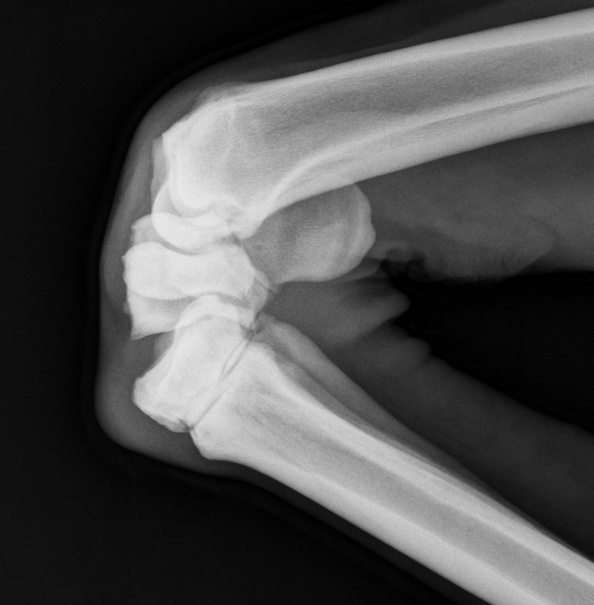
Some key points to remember about X-rays. Remember that X-rays are stopped differently by different density tissues. Bone, which is very dense, does not allow many X-rays to pass through it, and results in a white area on the image. On the other hand, air stops no X-ray at all, and when X-rays pass through the air and hit the sensor, what you see on the image is black.
Limitation #1 – The problem is that, just like the air, most of the non-bone tissues of the horse don’t stop many X-rays, either. Things like tendons and ligaments and blood vessels end up looking pretty black, too. That’s why, for example, when your horse has a tendon injury, if you want to see what’s going on, we need to use a different imaging technique, such as ultrasound.
Take Home Message #1 – If you want to look at bone, X-rays are a good choice. If you want to look at something else, use something else.
Limitation #2 – X-rays can not predict the future. X-rays can’t tell you how fast a known problem will get worse and they also can’t tell you if a “change” is going to develop into something significant in a horse that is currently sound.
Take Home Message #2 – You have to ride the horse; you can’t ride the X-ray.